Cam Impingement
Abnormal contact between the proximal femur (ball side of the joint) and acetabulum (cup side of the joint) may result from a variety of conditions. In some cases, it is genetically determined that you will have the problem. In some cases, you may have a previous fracture in the area of the hip ball. Chronic impingement may cause damage to the cartilage as a result of excessive contact with the abnormally shaped parts of the hip.
CAM impingement occurs when a nonspherical (not round) femoral head-neck rotates in the acetabulum. This mechanism defines a mismatch between the femoral neck and the acetabulum (cup side of the joint. There is often a bone lesion or subtle prominence on the superior femoral neck on xrays that your doctor may get. On the right, below you can see that the ball has an indentation (at the red arrow), on the left the ball has an area (red arrow) that makes it more convex than concave, causing the mismatch of the cup and the ball.


With the use of a diagram, the process that occurs in the hip joint is more obvious, as seen below. On the left, is the femoral head starting to go into the socket side of the joint. The yellow area represents the area of extra bone that has developed and is starting to engage the cartilage (red area). On the right, as the hip is brought into the joint, the yellow area is seen to further run into the cartilage (red) ultimately causing damage.

Over many repetitions, the cartilage is damaged and this eventually leads to a complete disruption of the smooth surface of the joint.
Imaging
Standard xrays best show the shape of the femoral head and neck and that is the first study that should be ordered by your physician. A true AP pelvis, AP and lateral of the affected hip highlight structural landmarks allowing assessment of head-neck offset and evidence of cam impingement.
There are several measurements that are made, including whether your joint space is adequate for a hip arthroscopy (you need at least 2 mm of space to have a good outcome with arthroscopy). In addition, an angle that is called the alpha angle is used to measure the degree of cam lesion that is evident. In most cases, an angle over 50 degrees is thought to be abnormal and likely requires surgical intervention. In the images below, the right side is a normal angle of 30 degrees. On the left, the area of extra bone can be seen outside of the circle and there is a very high alpha angle.

Further diagnostic imaging can document intra-articular pathology and assist in preoperative planning. MRI arthrograms are capable of detecting labral and less reliably, cartilage (chondral) injuries. An arthrogram involves injecting a small amount of dye into the joint so that any tears that are present are better seen with the MRI study that is done. At the same time as the injection is placed in the joint for the dye, your doctor may order a cortisone injection to help alleviate some of the pain associated with the tearing that is present and also to serve as an indicator that your pain is truly coming from the hip joint and not somewhere else, such as you back.
Asymmetric fluid presence surrounding the femoral neck on T2 images suggests inflammation in the peripheral compartment. The arthrogram will also define the three dimensional anatomy of the femoral head-neck offset, herniation pits, paralabral cysts, and ossification of the acetabular rim. In the example on the left below, the patient has a large cam lesion (by the red arrow), while there is a labral tear in the posterior (back area) of the hip, as seen at the white arrow. On the right, there is a labral tear with a chondral injury that is well seen.


A CT scan with 3D image reconstruction may also be used for preoperative planning, especially if treating complex problems or failed surgeries. The 3D CT scan offers a thorough analysis of a cam or pincer lesion, especially for those that are believed to extend posterior on plain radiographs. Below, is an image of a 3D CT showing a small cam lesion (arrow) that was not seen on xrays.
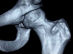
3D CT scan showing a cam lesion over the front to the hip
Treatment
Treating pincer lesions from acetabular retroversion is addressed arthroscopically with a cam resection and in most cases, a labral repair. The first part of the procedure fixes any of the labral tearing or cartilage damage as discussed above. Following the reattachment of the labrum the bone reshaping is performed with the use of both the arthroscope and x-ray at the time of the surgery.
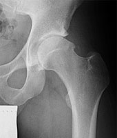
Pre-op Radiographs of a Cam Resection
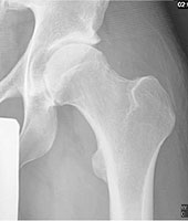
Post- op Radiographs of a Cam Resection